1
FDA 510(k)-cleared base
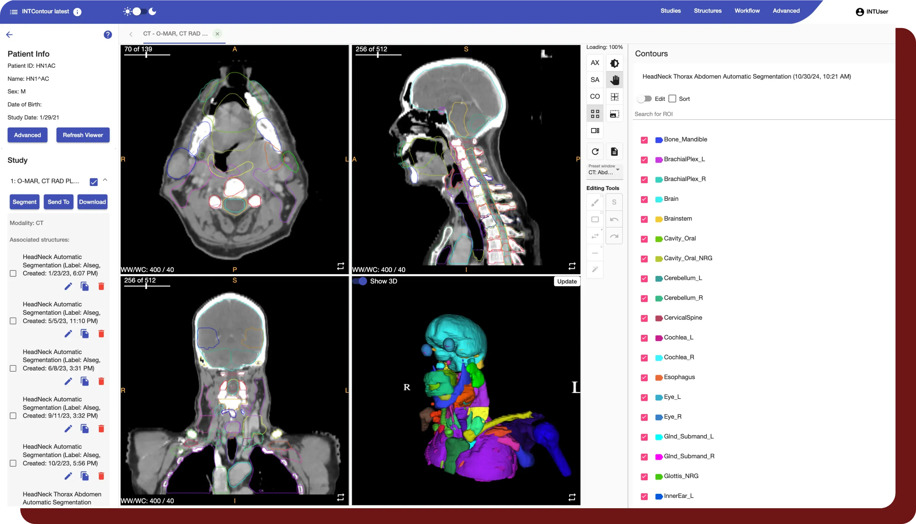
Ready to deploy on day one. 120+ OARs across CT, MRI, and PET/CT. Eclipse TPS integration, stand-alone web, automated DICOM workflows.
INTContour · AI-Native Auto-Contouring Platform
INTContour ships with FDA 510(k)-cleared models covering 120+ OARs and lymph node groups across CT, MRI, and PET/CT. On top of that, your team can train custom segmentation models on your own institutional data — the capability the Mayo Clinic study used to outperform vendor baselines on its own protocols.
Why teams pick INTContour
1
Ready to deploy on day one. 120+ OARs across CT, MRI, and PET/CT. Eclipse TPS integration, stand-alone web, automated DICOM workflows.
2
When a vendor model doesn't fit your protocol — iodinated spacers, rectal balloons, custom labels — your team retrains on local data without writing code. Mayo Clinic proof point ↓
3
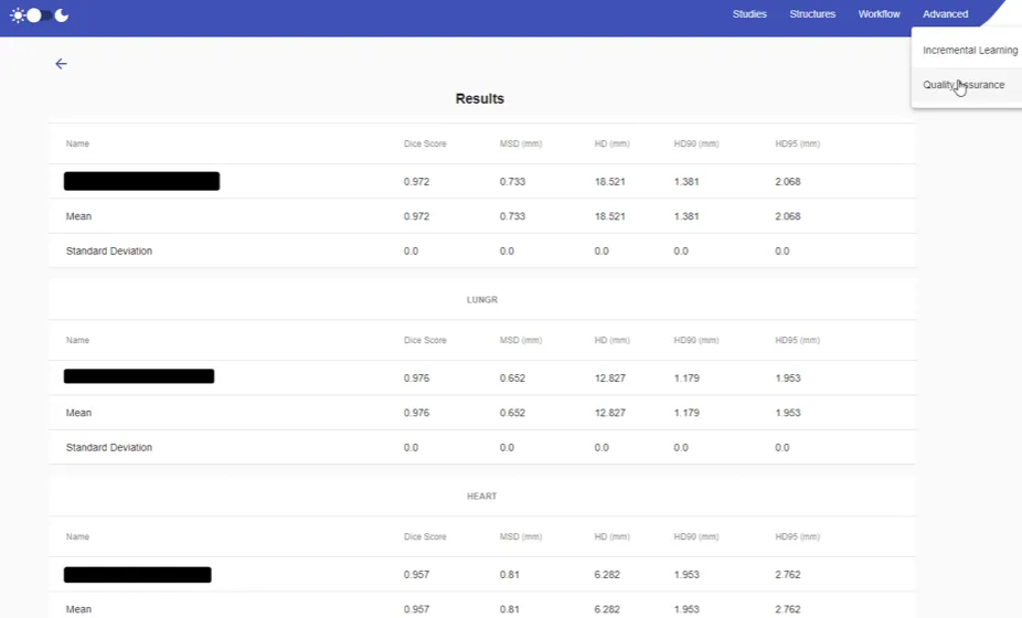
Dice, Mean Surface Distance, and Hausdorff Distance against ground truth — built into the same tool that produced the contour.
Available segmentations
Approved structures plus investigational-use structures across the four major regions covered today.
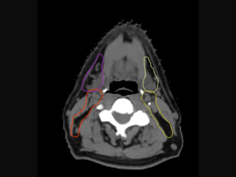
Mandible, Brachial Plexus, Brain, Brainstem, Cochlea, Eye, Lacrimal Gland, Submandibular Gland, Larynx, Optic Chiasm, Optic Nerve, Parotid, Pharynx, Pituitary, and more. Investigational: GTV Lymph Node, GTV Primary Tumor, Cerebellum, Neck Levels Ib/II/III/IVa, NRG-standard variants.
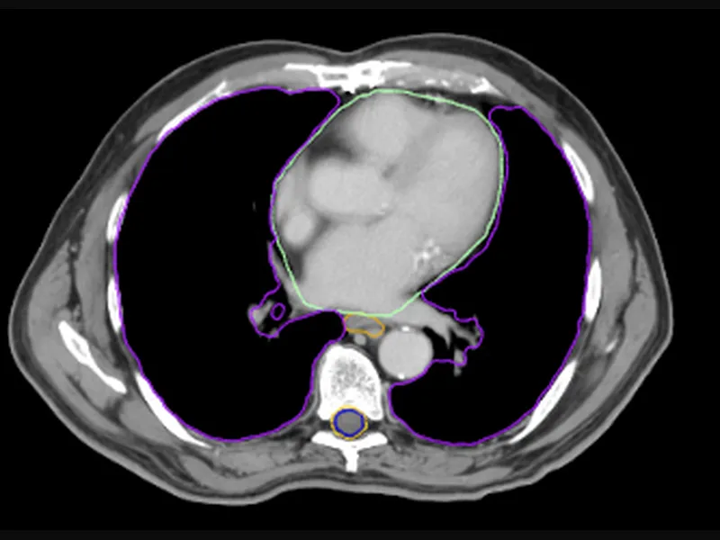
Lung, heart and cardiac substructures, esophagus, spinal cord, and chest-wall structures.
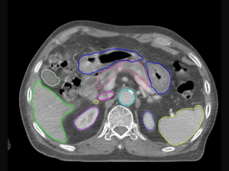
Liver, kidneys, stomach, duodenum, pancreas, bowel, spleen, and adrenal glands.
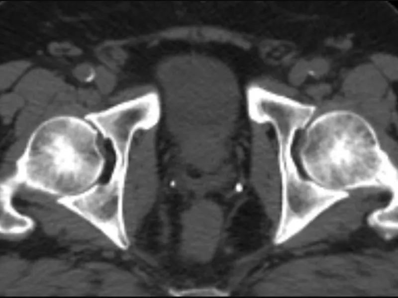
Male: prostate, seminal vesicles, bladder, rectum, femur heads, penile bulb, urethra. Female: uterus, cervix, ovaries, bladder, rectum, vagina, sigmoid, bowel bag.
Robust quality assurance
INTContour automatically generates quality assurance reports based on user selections to validate segmentation. Calculate Dice Score, Mean Surface Distance, and Hausdorff Distance between AI outputs and ground truth values with a single click.
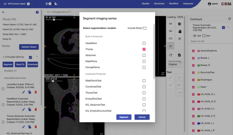
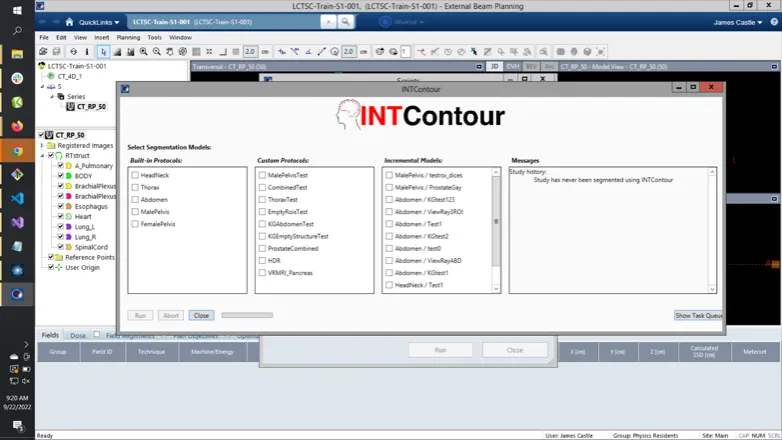
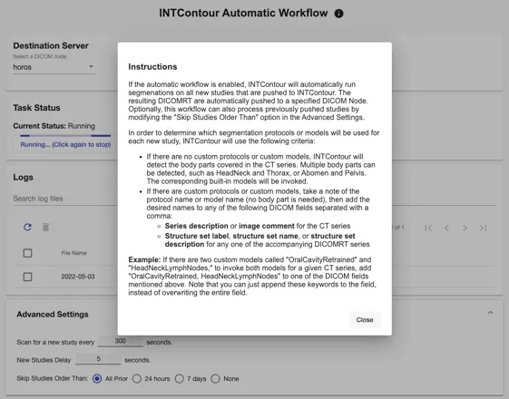
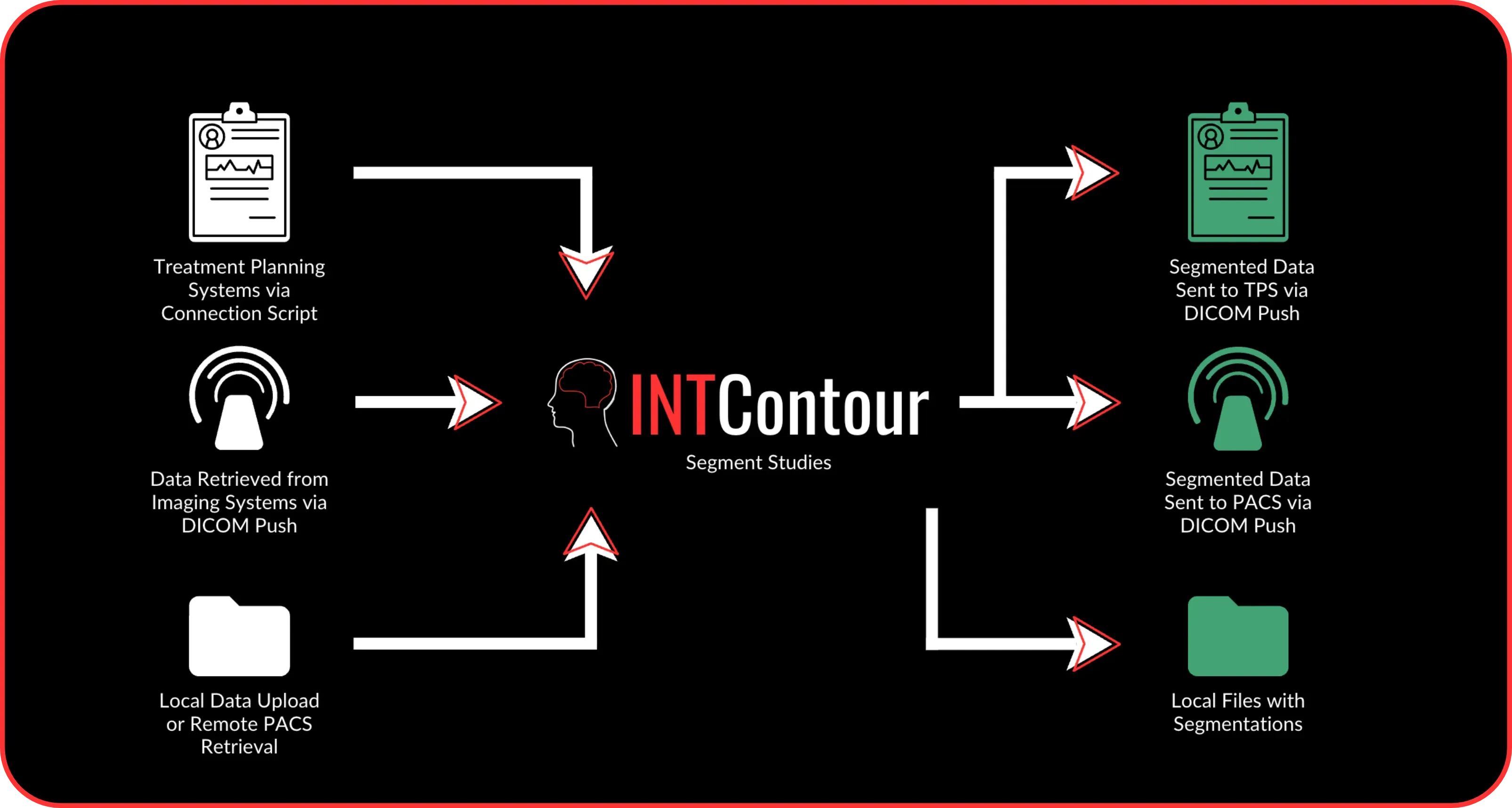
Powerful workflow connectivity
INTContour operates as a stand-alone web-based system, or integrates with applications via Automatic Workflow or TPS Connectivity. Using Automatic Workflow, INTContour efficiently segments the desired studies and transfers them back to the main application in use.
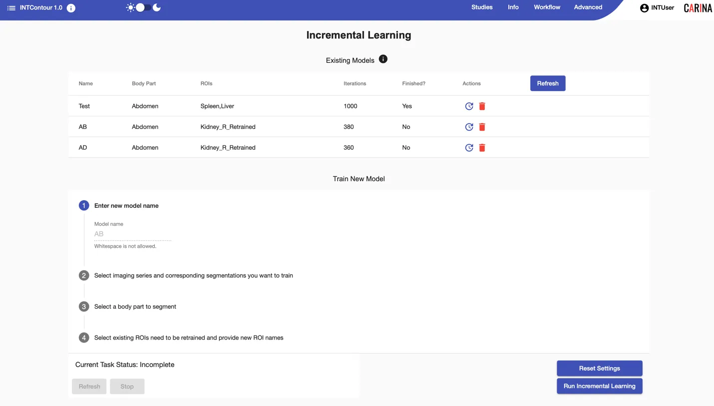
Research tools
Streamline your research with intuitive features to train models without coding and process large volumes of studies with a single command using award-winning AI algorithms.
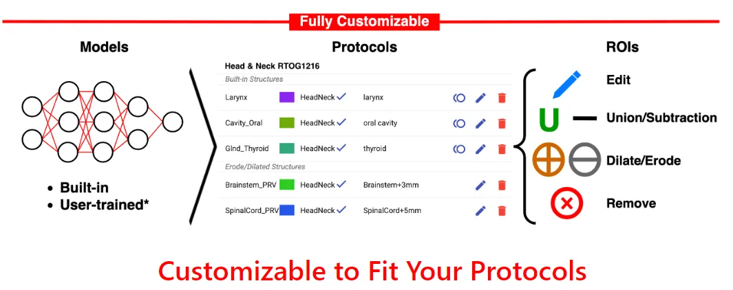
* Tools provided as a research-use feature. INTContour does not assure the accuracy and performance of newly trained models.
Award-winning algorithm lineage
Proof point · Mayo Clinic
The strongest evidence we have for incremental learning.
Setting. Prostate planning with iodinated spacer and rectal balloon — anatomy not represented in vendor training data. Baseline. Three major vendors' built-in deep-learning auto-segmentation models underperformed on prostate, seminal vesicles, bladder, rectum, and femur heads. Intervention. INTContour incremental retraining on 100 cases. Readout. Blinded 5-point rating by 6 GU radiation oncologists plus 2 RO residents, 115 test cases.
≥ manual in 50%+
of cases across every group and reviewer — range 63.8% (SV, junior resident) to 100% (bladder, RO1).
Duan et al., Medical Physics, 2023.
Duan et al., Radiotherapy & Oncology, 2025.

Research collaborations
Publications
Selected INTContour-related publications. See the full list in Resources.
30-minute walkthrough with our scientific team.